More than 3 million Americans experience arrhythmia - the heart beats too fast, too slow, or too irregularly. One of the irregular heartbeat form - ventricular tachycardia - is the leading cause of sudden cardiac arrest.
Irregular heartbeat or arrhythmia can interrupt the normal flow of blood and put you at risk of blood clots and stroke. An implanted defibrillator can help reboot the heart.
Clarence Mankin 74, a retired Lutheran pastor in Atlanta, Illinois, has served in the US Army during the Vietnam War. He suffered his first heart attack at 44, which he attributed to Agent Orange exposure.
The once very active Mankin - who could easily walk 10,000 steps a day, started getting jolts all the time. At one point, he had even 11 episodes a day which forced him from doing anything. He stopped walking, driving, or even leave his couch.
When Mankin experiences sudden irregular heartbeat, he would pass out. The implanted defibrillator would reboot his heart. Then, he would wake up dazed but alive. Once his doctor adjusts the defibrillator sensitivity, it would shock his heart and he would faint. The symptoms before his blackout are dizziness and lightheadedness. Mankin would know to brace himself for the jolt.
Mankin says, "It's like a good thump in the chest. It feels like you are going to die. I'd try to grab hold of something, or find something to sit down on, which occasionally I was able to do."
Ventricular tachycardia (VT)
An abnormal rapid heartbeat may result from other cardiac problems. A heart attack may injure the heart and can cause some cells to die which leads to scar tissue.
When there is any scar or damage to the cells, it may not function as is supposed to, which causes the heart to short-circuit. This may cause erratic heartbeat and the blood flow throughout the body may be interrupted.
Treatments include an implantable cardioverter-defibrillator or ICD below the collarbone under the skin. It helps the heart to beat normally. Another option is called catheter ablation in which doctors use heat to destroy abnormal heart tissue.
Catheter ablation is an invasive procedure where the doctors use a fine tube through the femoral vein up into the heart. An electrode tip selectively burns away the damaged heart cells. This procedure can last anywhere between four to 10 hours and the recovery period is long. Even with the latest technologies, the physicians try and ensure to destroy the damaged heart cells, but this treatment may not work always. The side effects of this treatment range from infections to bleeding problems.
In a 2016 study, patients undergone catheter ablation procedure saw their tachycardias return. Same was the case with Mankin.
Revolutionary Treatment
The collaboration between a School of Medicine cardiologist and a radiation oncologist has led to a revolutionary treatment.
And, Mankin is one of the earliest patients to undergo the treatment. A beam of radiation will be given directly to the heart. Mankin's cardiologist referred him to a heart rhythm specialist Dr. Phillip S. Cuculich, an associate professor of medicine at Washington University.
Cuculich teamed up with Cliff G. Robinson, MD, also an associate professor of radiation oncology to provide a revolutionary arrhythmia treatment. This new treatment is like a game changer for patients with fewer options.
How did they test?
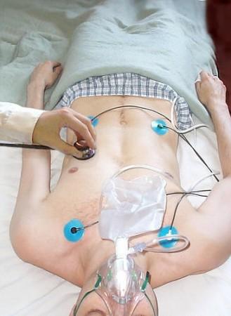
Mankin to undergo the procedure had to don a specially designed vest covered in 252 electrodes (12 for a typical electrocardiogram). Mankin was briefly induced with VT using his defibrillator. A panoramic map was produced while Mankin lay inside a cylindrical chamber. A custom mold held Mankin in place which prevented from moving or breathing deeply.
Robinson used the panoramic map to direct an intense, focused beam of radiation at his heart. The high-energy radiation beam is typically used to blast cancer cells. The procedure takes about 15 minutes or lesser. The particle stream intentionally zapped the malfunctioning cells.
Robinson said, "I've spent my entire life as a trainee and an attending physician thinking about ways to avoid dosing healthy tissues. But this is already an injured region. If you think about it as a diseased part, where Phil would have no problem going in with a catheter to burn that area, well, we are doing the same thing. There are other ways to get energy inside the body."
The whole radiation therapy was done while Mankin was fully conscious at Barnes-Jewish Hospital on September 8. He was able to walk out of the hospital immediately after the procedure without any pain.
Surprising follow-up results
Mankin had to return again to the hospital for a follow-up. In January, Cuculich downloaded data from his Mankin's defibrillator which tracks the jolts that he feels.
After he saw the device history, Robinson was surprised at the results. Prior to the procedure, the results showed in the graph resembled a forest with so many spikes of jolts. After September 8 and months after Mankin's procedure, the graph was clear and the device had not activated at all and the tachycardia was gone.
Mankin has not had a single jolt from his defibrillator and is now able to walk 5000 steps a day.
Mankin said, "It's hard to describe. Now when I walk, I don't feel like something terrible is going to happen to me," Mankin said. "I don't know if this will extend my life or not, but it has made the quality of my life much better."
Many patients enrolled in the clinical trial of Cuculich and Robinson. Their study results were published in The New England Journal of Medicine.
The trial is clearly a winner. As in the first tests, the five patients collectively experienced 6577 episodes of VT in the three months prior to their radiation procedure. In the nine months afterward, they experienced only four.
How did the treatment evolve?
Cuculich spent many years in refining techniques to map the heart and the precise location that is damaged. The technique was developed by the Fred Saigh Distinguished Professor of Engineering, director of the Washington University Cardiac Bioelectricity and Arrhythmia Center, and Cuculich's mentor - Yoram Rudy, Ph.D.
Cuculich says a detailed image of heart dysfunction was not enough.
He said, "Even if you could non-invasively image the heart, we still required entry into the body with a catheter to fix it," Cuculich said. "It became clear that the place to make an impact was to non-invasively treat it."
Cuculich met Robinson through neurosurgeon Albert H. Kim, MD, Ph.D. At this point, Robinson was also looking for innovative ways to use stereotactic radiation, a precise, high-energy dose of radiation, beyond cancer treatment.
Robinson said, "When we first met to discuss this, I was really concerned about how to hit a 5 mm area in a moving, beating heart. But Phil's first question was, 'How big of an area can you treat?' And I said, 'This is going to be a good friendship.' Because as radiation oncologists, we don't treat parts of tumors. We treat the whole kit and caboodle."
Cuculich and Robinson discussions
From many discussions that they had regarding each other's specialties and their patients, they learned more about themselves.
Robinson said, "That was one of the keys to this initial success, that we came together and talked about it. That this could happen is one of the unique things about being at Washington University."
They developed a technique that relies on multiple imaging methods - MRI, CT and PET scans which combine with the electrode vest to produce a detailed map of the heart and precisely locate where the arrhythmias are coming from. The imaging methods involved patients taking several hours, occasionally spread over multiple days.
"Then Cliff and I sit down together and collaborate over this information, and we come to a conclusion about where the scar is, so we are as precise as possible. We want to avoid any structures that are important to the heart, and keep the potentially damaging energy within the scar," Cuculich said.
Robinson said that the procedure is much like cancer treatment - to know how deep to go, how much tissue to take and leave. In some patients like Mankin, they have large areas to be treated and in some, the cells deep within the heart muscle that cause the heart to beat out of sync are not reachable with the tiny catheter tip.
So, the radiation beam can penetrate deeper, target all the damaged cells.
The physicians' concern over some patients resistance against the fearful word 'radiation' proved wrong. They said that their patients have been less resistant than they expected because they were desperate for any solution.
Risks of radiation
With such high-energy beam radiating a very small site, there are risks involved. Toxicity, effects on the surrounding tissues and cells are some side effects of radiation. Robinson said, patients from the initial trial and the ongoing study have not reported any complications or pulmonary symptoms due to radiation.
While some did experience mild inflammation in the lung adjacent to the target, it was treated within a year. Still, the potential long-term ill effects of the radiation dose are unknown.
Roy John, Ph.D., professor of medicine at Vanderbilt University Medical Center and Stevenson who co-authored the editorial explaining of the procedure's potential in the journal said, "If this is to be used in younger, less-ill patients, the issue of collateral damage will become more relevant."
Both Robinson and Cuculich say that the early results are promising. They are finalizing their results from the ENCORE-VT trial, which included 18 patients along with Mankin.
Robinson said, "We have created an entirely non-invasive process to map and treat arrhythmia, and we can do it in less than 10 minutes. This approach can fundamentally change the way we approach these heart rhythm problems."
Radiation procedures can become complementary rather than complete replacement of other therapies. It provides as another option for VT patients who have no other options for treatment and are facing survival rates below 20 percent.


