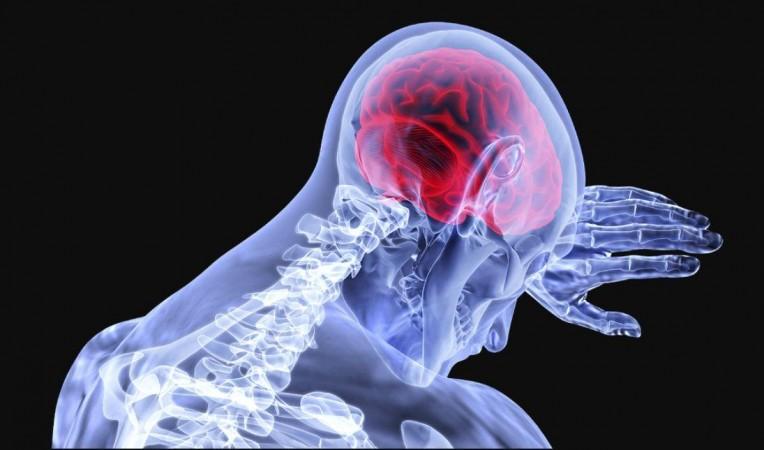
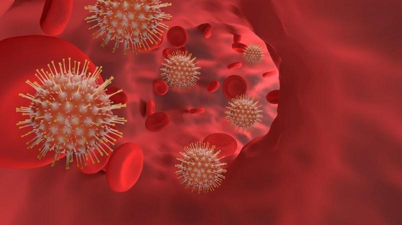
Despite being a respiratory virus, the SARS-CoV-2 can wreak havoc on one's nervous system. Not only have millions of individuals across the world reported neurological symptoms while enduring the COVID-19 infection, but also suffering from them after recovering from the disease. Now, offering new insights into the effect of the virus on the brain, scientists have found mechanisms associated with how the novel coronavirus infection affects the organ.
According to a new multi-institutional study, COVID-19 patients struggling with severe neurological symptoms exhibit signs of impairment in the blood-brain barrier, a semipermeable membrane separating the cerebrospinal fluid (CSF) from the blood. It was also found that there were changes in the structure of their brains, commensurate to the severity of their symptoms. The findings were published in the journal Nature Communications.
"In our study, we show how coronavirus can affect the brain. The virus triggers such a strong inflammatory response in the body that it spills over to the central nervous system. This can disrupt the cellular integrity of the brain," said Dr. Gregor Hutter, lead author of the study, in a statement.
Examining Neurological Effects of COVID-19

Some of the most common symptoms during the course of the COVID-19 infection are loss of sense of smell (anosmia) and taste (ageusia), both of which are neurological in nature. Despite recovering from the disease, numerous survivors continue to battle "long COVID": a set of lingering symptoms extending beyond 12 weeks after infection.
Among these symptoms are neurological ones such as memory lapses and inability to concentrate (brain fog)–also known as "neuro-Covid"–that vary in severity across different groups of individuals. Through the current study, the researchers aimed to examine the blood plasma and CSF of affected patients, and probe how varying severities of neuro-COVID can be detected and predicted through it.
For the study, the team enrolled 40 patients with COVID–19, who were experiencing varying degrees of neurological symptoms. Blood plasma and cerebrospinal fluid (CSF) samples were collected from the participants and compared with that of healthy individuals (control group) in order to ascertain changes associated with neuro-COVID.
The patients' brain structures were also evaluated using techniques such as computed tomography (CT) and magnetic resonance imaging (MRI). In order to investigate long-COVID symptoms, the participants were subjected to a follow-up 13 months after the contraction of the infection.
Damage to the Blood-brain Barrier

The team identified a link between excessive immune response and neurological symptoms among patients in whom the symptoms were severe. It was found that these participants exhibited prominent signs of blood-brain barrier impairment, manifesting as hole-like formations.
According to the authors, this could potentially be the result of a "cytokine storm"–an erratic immune response of the body to infections. Another finding was that the individuals' antibodies targeted portions of the body's own cells–indications of an autoimmune reaction–owing to the unrestricted immune response.
Explaining these observations, Dr. Hutter said: "We suspect that these antibodies cross the porous blood-brain barrier into the brain, where they cause damage." It was also learnt that there was an uncontrolled activation of microglia, specialised immune cells found in the brain, in participants experiencing severe neurological symptoms.
Changes in the Structure of the Brain

Next, the researchers examined whether the brain structures of the enrollees showed any perceptible signs of the severity of their neurological symptoms. It was observed that in participants with acute neuro-COVID symptoms, the brain volume–in comparison to healthy individuals–was lower at specific locations, particularly in the area of the brain that regulates smell, the olfactory cortex.
"We were able to link the signature of certain molecules in the blood and cerebrospinal fluid to an overwhelming immune response in the brain and reduced brain volume in certain areas, as well as neurological symptoms," noted Dr. Hutter. He also emphasised the need to study these biomarkers in larger groups of participants and the development of a blood test that can predict the seriousness of the infection, including potential long COVID and neuro-COVID, on its onset.
According to the team, the identified biomarkers may serve as potential targets for drugs that focus on the prevention of the damage resulting due to COVID-19 infection. MCP-3, a cytokine involved in the excessive immune response, is one such biomarker, and Dr. Hutter stated that there is medicinal potential to inhibit the protein.


