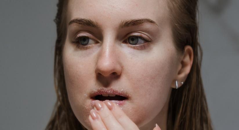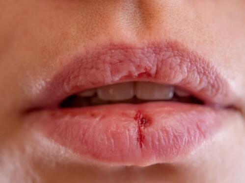
Swiss scientists have successfully created 3D cell models using lip cells. This achievement, a first in the world, could potentially revolutionize the treatment of lip injuries and infections. Lip cells, which behave differently from other skin cells, have not been used in models until now. Dr. Martin Degen, from the University of Bern in Switzerland, emphasized the importance of this development, stating, The lip is a very prominent feature of our face. Any defects in this tissue can be highly disfiguring. But until now, human lip cell models for developing treatments were lacking.
To address this gap, the scientists immortalized donated lip cells, allowing them to develop clinically relevant lip models in the lab. The cells were donated by two patients, one undergoing treatment for a lip laceration and the other for a cleft lip. The team used a retroviral vector to deactivate a gene that halts a cell's life cycle and altered the length of the telomeres on the ends of each chromosome to improve the cells' longevity.
The new cell lines were then rigorously tested to ensure they retained the same characteristics as primary cells and to check for any chromosomal abnormalities. The scientists then conducted tests to see how the cells might perform as future experimental models for lip healing or infections. They scratched samples of the cells to see if they could act as accurate proxies for wound healing. The results showed that untreated cells closed the wound in eight hours, while those treated with growth factors closed the wound more quickly. These results were consistent with those seen in skin cells from other body parts.

The team then developed 3D models using the cells and infected them with Candida albicans, a yeast that can cause serious infections in people with weak immune systems or cleft lips. The cells performed as expected, and the pathogen rapidly invaded the model as it would infect real lip tissue. Dr. Degen expressed optimism about the potential applications of these 3D models, stating, We are convinced that 3D models established from healthy immortalised lip cells have the potential to be very useful in many other fields of medicine.
This development is particularly significant given the complexity of craniofacial development, which involves the orchestrated interactions of multiple cell types. Neural crest cells (NCCs) are a critical population in this process. Errors in the complex, spatio-temporally regulated networks of signaling cues can result in a variety of craniofacial anomalies (CFAs).
CFAs can appear in isolation, as part of a syndrome with defects due to mutations in single genes or chromosomal abnormalities, or in combination with other defects without an identified genetic background. CFAs can have a significant impact on a child's self-perception and social integration due to esthetical concerns, representing a high economic, social, and psychological burden for affected individuals, their families, and society as a whole.
The development of 3D cell models using lip cells could potentially contribute to a better understanding of CFAs and optimize treatment options tailored to specific patient needs. In a related development, a new experimental pipeline has been developed to study membrane breaching events during C. albicans epithelial cell invasion at the single cell level. This pipeline could potentially provide valuable insights into the mechanisms of C. albicans invasion and contribute to the development of more effective treatments for infections caused by this pathogen.


